Faça uma busca em todo o nosso site.
Pesquisas e Artigos em Odontologia.
Pesquisas e Artigos em Estética
Pesquisas e Artigos em Fisioterapia
Pesquisas e Artigos em Medicina
Pesquisas e Artigos em Engermagem
Pesquisas e Artigos em Podologia
Pesquisas e Artigos em Fonoaudiologia
Pesquisas e Artigos em Laserpuntura
Pesquisas e Artigos em Veterinária
O Instituto Nupen é uma fundação criada para permitir e contemplar a ampliação do escopo de atuação inicial do Núcleo de Pesquisa e Ensino de Fototerapia nas Ciências da Saúde.
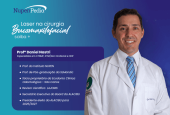
A biofotônica tem evoluído constantemente, uma grande novidade é o laser azul cirúrgico de 450nm, com altíssima afini...
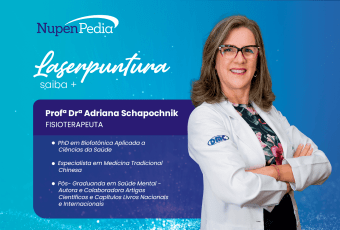
A Laserpuntura consiste na irradiação do laser de baixa intensidade nos pontos sistêmicos e auriculares no regime ope...
O mês de dezembro marca o início da campanha “Dezembro Vermelho”, instituída no Brasil pela Lei nº 13.504/2017 que ch...